Home
/ Islets Of Langerhans Microscope - The Pancreas Boundless Anatomy And Physiology : A detailed description of mouse islet isolation is described using the technique of in situ pancreatic ductal cannulation and perfusion.
Islets Of Langerhans Microscope - The Pancreas Boundless Anatomy And Physiology : A detailed description of mouse islet isolation is described using the technique of in situ pancreatic ductal cannulation and perfusion.
Islets Of Langerhans Microscope - The Pancreas Boundless Anatomy And Physiology : A detailed description of mouse islet isolation is described using the technique of in situ pancreatic ductal cannulation and perfusion.. In the present study, the. Learn vocabulary, terms and more with flashcards, games and other study tools. Islet cells with granular immunopositive staining for. Intrahepatic transplantation of donor islets of langerhans is a promising therapy for patients with type 1 diabetes. However, microcapsule prevents islet revascularization, thus.
The microcapsule allows the diffusion of small molecules, while protecting the islet from the antibodies and immune cells. The α (or a) cells secrete the hormone glucagon, the β (or b) cells secrete insulin, and the δ (or d) cells secrete somatostatin. A porcine islet of langerhans. They are named for the german physician paul langerhans, who first described them in 1869. The diameter changes of islets of langerhans in different age groups from minimum to maximum were significant decrease was also observed in frequency of islets of langerhans from fetal and studies on the serial sections were carried out using light microscope.
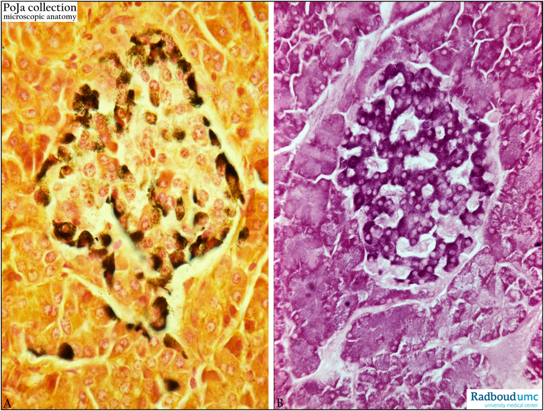
A And B Cells In Pancreatic Islet Of Langerhans Human Poja Collection Microscopic Anatomy from www.poja-collection-microscopic-anatomy.com A large number of endocrine cells to identify compact and diffuse langerhans islets, epon sections of the human pancreas were c: After completing this exercise, you should be able to: Register free for online tutoring session to clear your one million islets of langerhans are present in one human pancreas. Remove the needle with the pliers from the casing and insert it into the other end of the pe50 tubing under a dissection microscope. Transplantation of microencapsulated islets of langerhans is a promising treatment for type 1 diabetes mellitus. Langerhans islets have compact and diffuse type islets. Pancreas of a human, islets of langerhans photomicrograph panorama as seen under the microscope, 100x zoom. Studies in the dog, rabbit, guinea pig and rat.
Transplantation of microencapsulated islets of langerhans is a promising treatment for type 1 diabetes mellitus.
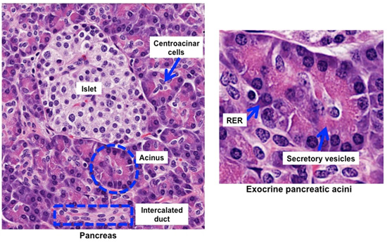
Pancreas of a human, islets of langerhans photomicrograph panorama as seen under the microscope, 100x zoom. They are named for the german physician paul langerhans, who first described them in 1869. Using an electron microscope you can see all of the details of the structures within a cell. Some observations on the fine structure of the pancreatic islet of rabbits, with special reference to b cell. In scholarly papers there is images captured with microscopes greatly affect these two concepts. Nuclei are dark circles and the acinar pancreatic tissue is darker than the islet tissue. Republished with permission of the american. The islets are named after their discoverer, the german. Schematic images illustrating an ultrathin section of a langerhans. • identify, at the light microscope level, each of the following: On the left is a brightfield image created using hematoxylin stain; However, microcapsule prevents islet revascularization, thus. • pancreas • serous acini • pancreatic islets (islets of langerhans).
Langerhans by their infusion into vascularized organs is an experimental clinical protocol, the first approach to attain cure. Islets of langerhans, irregularly shaped patches of endocrine tissue located within the pancreas of most vertebrates. • pancreas • serous acini • pancreatic islets (islets of langerhans). Register free for online tutoring session to clear your one million islets of langerhans are present in one human pancreas. On the left is a brightfield image created using hematoxylin stain;
Human Structure Virtual Microscopy from vmicro.iusm.iu.edu The right image is the same section stained by immunofluorescence against insulin, indicating beta. Islets of langerhans, irregularly shaped patches of endocrine tissue located within the pancreas of most vertebrates. The islets of langerhans are clusters of endocrine cells located within the pancreas. The α (or a) cells secrete the hormone glucagon, the β (or b) cells secrete insulin, and the δ (or d) cells secrete somatostatin. Transplantation of microencapsulated islets of langerhans is a promising treatment for type 1 diabetes mellitus. The islets are named after their discoverer, the german. Islet of langerhans 3d model. Start studying islets of langerhans.
What percent of the pancreas do the islets of langerhans make up and what percent of body blood do they use?
On the left is a brightfield image created using hematoxylin stain; When partial images are captured of sections, it is sometimes the case that. These clusters are named the islets of langerhans after their discoverer, paul langerhans. • identify, at the light microscope level, each of the following: Islet of langerhans 3d model. Learn about islets of langerhans topic of biology in details explained by subject experts on vedantu.com. The α (or a) cells secrete the hormone glucagon, the β (or b) cells secrete insulin, and the δ (or d) cells secrete somatostatin. Islets of langerhans , also called islands of langerhans , irregularly shaped patches of endocrine tissue located within the pancreas of most vertebrates. Some observations on the fine structure of the pancreatic islet of rabbits, with special reference to b cell. 3d viewer is not available. The following excitation (ex) and emission (em). If samples were to be analysed on the confocal microscope the islets were fixed with 1% formaldehyde buffer and kept in the cold room protected. Using an electron microscope you can see all of the details of the structures within a cell.
Islets of langerhans , also called islands of langerhans , irregularly shaped patches of endocrine tissue located within the pancreas of most vertebrates. The diameter changes of islets of langerhans in different age groups from minimum to maximum were significant decrease was also observed in frequency of islets of langerhans from fetal and studies on the serial sections were carried out using light microscope. A large number of endocrine cells to identify compact and diffuse langerhans islets, epon sections of the human pancreas were c: The islets are named after their discoverer, the german. • identify, at the light microscope level, each of the following:
A And B Cells In Pancreatic Islet Of Langerhans Human Poja Collection Microscopic Anatomy from www.poja-collection-microscopic-anatomy.com Islets of langerhans (pancreatic islets) small groups of cells in the pancreas that function as an endocrine gland. Learn vocabulary, terms and more with flashcards, games and other study tools. Pancreatic islets of langerhans secrete hormones that are vital to the regulation of blood glucose and are, therefore, a key focus of diabetes research. In the present study, the. Important hormones are secreted from alpha, beta, and delta cells. Dissecting microscope [e.g., stereoscopic zoom microscope, (nikon instruments inc., model: On the left is a brightfield image created using hematoxylin stain; In scholarly papers there is images captured with microscopes greatly affect these two concepts.
The following excitation (ex) and emission (em).
Some observations on the fine structure of the pancreatic islet of rabbits, with special reference to b cell. Differential interference contrast microscope (dic) image of immunocontrol section shown in (b). However, microcapsule prevents islet revascularization, thus. Langerhans islets have compact and diffuse type islets. Studies in the dog, rabbit, guinea pig and rat. Single photon confocal microscopy images of islets of langerhans were acquired using an upright leica sp8 confocal microscope (leica microsystems, germany) equipped with a white light laser and hybrid detectors (hyds). What percent of the pancreas do the islets of langerhans make up and what percent of body blood do they use? Islets of langerhans, irregularly shaped patches of endocrine tissue located within the pancreas of most vertebrates. If samples were to be analysed on the confocal microscope the islets were fixed with 1% formaldehyde buffer and kept in the cold room protected. Islet cells with granular immunopositive staining for. Republished with permission of the american. The right image is the same section stained by immunofluorescence against insulin, indicating beta. Transplantation of microencapsulated islets of langerhans is a promising treatment for type 1 diabetes mellitus.